Fig. 8.

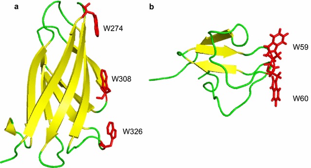
Crystal structures of CBM2 in Pyrococcus furiosus chitinase (PDB No. 2CWR, (a)) and CBM5 in Streptomyces griseus ChiC (PDB No. 2D49, (b)). The stick models of the Trp residues involved in binding carbohydrates are colored in red

Crystal structures of CBM2 in Pyrococcus furiosus chitinase (PDB No. 2CWR, (a)) and CBM5 in Streptomyces griseus ChiC (PDB No. 2D49, (b)). The stick models of the Trp residues involved in binding carbohydrates are colored in red