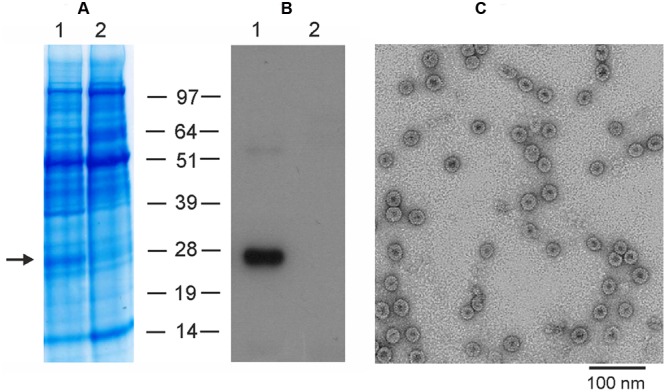
FIGURE 5.

Expression and purification of M2eHBc using the pEff vector. Coomassie brilliant blue stained gel (A) and Western blot (B) of proteins isolated from N. benthamiana leaves and separated by SDS-PAGE. M, molecular weight marker (kD); (1) total soluble proteins isolated from leaves infiltrated with vector pEff-M2eHBc; (2) total soluble proteins isolated from uninfiltrated leaves. Antibodies against M2e were used in Western blotting. (C) Transmission electron microscopy of plant-produced M2eHBc virus-like particles. The grid was negatively stained with 2% (w/v) uranyl acetate and the scale bar is shown.
