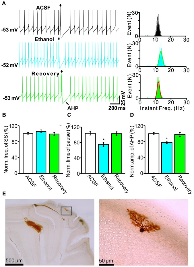
Figure 1.
Effects of ethanol on the spontaneous activity of Purkinje cells (PCs). (A) Left, representative traces showing the spontaneous activities of a PC in the presence of artificial cerebrospinal fluid (ACSF), ethanol (300 mM) and washout. Right, histograms show the instantaneous frequency of simple spikes (SS). (B,C) Bar graphs show the effect of ethanol on the frequency (B) and the pause (C) of SSs. (D) Summary of data showing the normalized amplitude of the afterhyperpolarization (AHP) in the presence of ACSF, ethanol (300 mM) and washout. (E) The photomicrographs show the morphology of the cell, which is shown in (A). The left column shows an overview of the location of the biocytin-labeled cell. The right column shows the detail of the biocytin-labeled cell. Note that ethanol significantly decreases both the amplitude of the AHP and the pause in SS firing. n = 8 cells per group. *P < 0.05, vs. ACSF.

