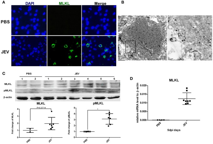
Figure 2.
MLKL mediated necroptosis is involved in JE. Mice were infected i.p. with PBS or JEV 5 × 10⁷ PFU in 200 μl PBS/20 g. At 5 dpi, brains were harvested for immunochemistry, immunoelectromicroscopy, westeron blot and qRT-PCR. (A) The immunochemistry of MLKL in the brain sections of PBS or JEV administered mice (x400). There was obvious staining of MLKL around the plasma membrane in the brain sections of JEV infected mice while it was invisible in control mice. (B) Immuno-electron microscopic study of MLKL. The right panels show magnified regions of the boxed areas. Arrows showed the membrane localization of MLKL. (C) Western-blotting (upper) and quantitation (down) of protein MLKL and pMLKL in PBS or JEV administrated group. There was increased expression of protein MLKL and pMLKL in JEV infected group (PBS = 2, JEV = 6, *P < 0.05). (D) The relative level of mRNA MLKL in PBS or JEV treated mice brain via qRT-PCR at 5 dpi. The data represents the relative mRNA level normalized with β-actin (PBS = 4, JEV = 8). The mRNA MLKL was significantly increased in JEV infected mouse brains compared with PBS group.

