
FIGURE 2.
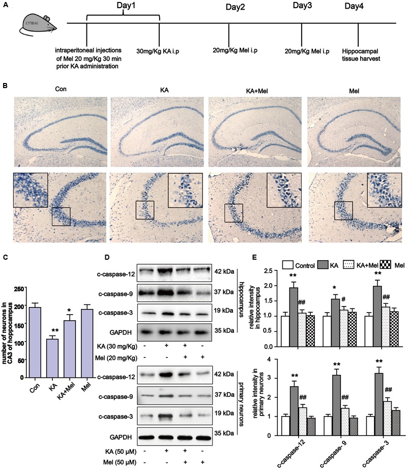
Melatonin reduces KA-induced neuron loss in vivo. (A) The administration procedure of melatonin and KA in C57 BL/6 mice. (B) Nissil’s staining assay was used to examine neuronal loss in KA and/or melatonin treated animals hippocampus tissue. Neuron numbers in hippocampus CA3 area were quantified in (C). (D,E) Expression levels of c-caspase-12, -9, -3 in hippocampal tissues and primary neurons. ∗P < 0.05, ∗∗P < 0.01, ∗∗∗P < 0.001 vs. controls; #P < 0.05, ##P < 0.01, ###P < 0.001 vs. the KA group; significant difference from the respective values determined by one-way analysis of variance test. n = 7 for Nissil’s staining; n = 3 for western blots.