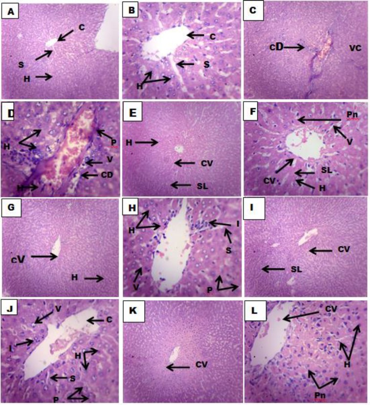
Figure 1.
Histologic sections of the liver treated with normal saline (10 ml/kg) A (x100) and B(x400), PCM (2.0g/kg) C (x100) and D (x400), silymarin (100 mg/kg) and PCM (2.0g/kg) E (x100) and F(x400), HL (250 mg/kg) and PCM (2.0g/kg) G (x100) and H (x400), HL(500 mg/kg) and PCM (2.0g/kg) I (x100) and J (x400, HL (750 mg/kg) and PCM (2.0g/kg) K (x100) and L (x400).
Keys: Central vein (CV) Sinusoidal lining (SL), Hepatic Artery (HA), Hepatic vein (HV), Hepatocytes (H) and Pyknotic nucleus (Pn), Cellular degeneration (CD), Vascular congestion (VC), Vascular degeneration (VD), Hepatocytic hyperplasia (H), Sinusoidal lining (SL), Vacuolation (V), Inflammation (I), Hepatocytic hyperplasia (HH), and Cellular proliferation (CP) .

