
Figure 6.
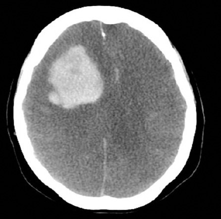
Brain computed tomography scan with contrast showed complete loss of gray and white matter differentiation of both cerebral hemispheres with large frontal hematoma, and complete effacement of extra axial cerebrospinal fluid spaces including lateral ventricles and basal cisterns.