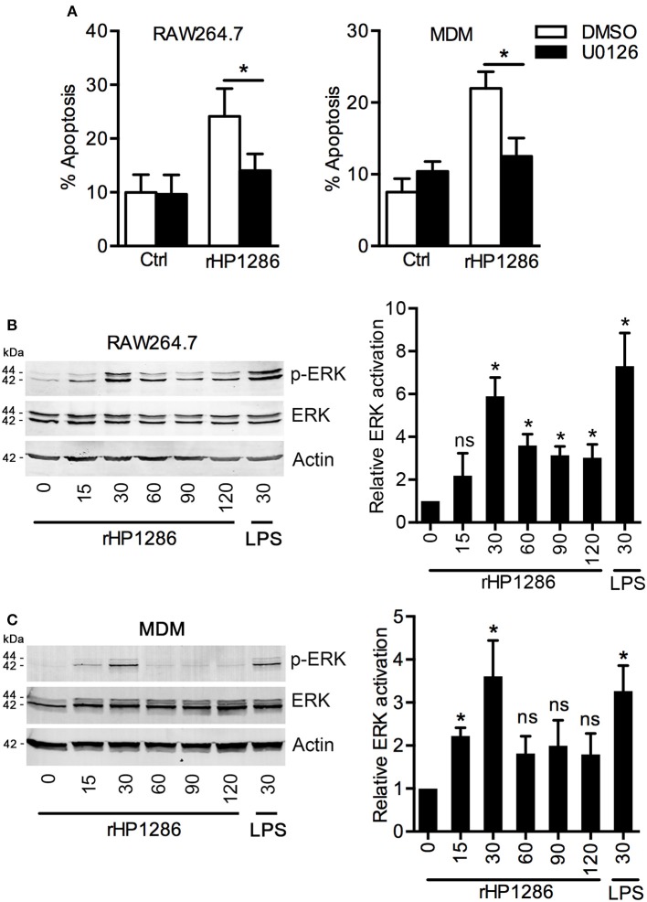
Figure 7.
The role of ERK MAPK in rHP1286-induced macrophage apoptosis. (A) RAW264.7 and MDM cells were pretreated either with vehicle control (DMSO) or U0126 (10 μM) for 30 min followed by treatment with rHP1286 (1 μg/ml). Apoptotic cells were analyzed by flow cytometry. The data shown is the mean ± SD of three independent experiments (RAW264.7) or three donors (MDM). Statistically significant differences are indicated by *(p < 0.05). RAW264.7 cells (B) or MDM (C) were treated with rHP1286 (1 μg/ml) for indicated time periods. As a positive control for ERK activation, RAW264.7 cells were treated with E. coli LPS for 30 min. Western blotting was performed with anti-phospho-ERK antibody. Blots were reprobed with anti-ERK and anti-actin antibody to ensure equal loading. Data shown is the representative of four independent experiments (RAW264.7) and three donors (MDM). The bar diagram indicates band intensities in western blots normalized to the actin control. *(p < 0.05) indicates statistically significant differences.

