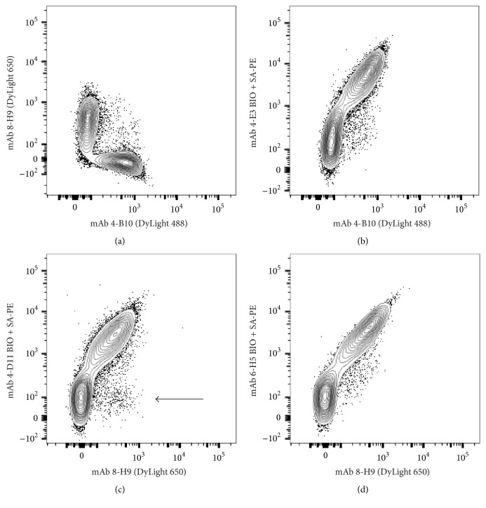
Figure 6.
Characterization of light chain reactive monoclonal antibodies. Mouse anti-ferret Ig mAb were used in combination for surface staining of ferret PBMC. Plots are pregated on B cell receptor expressing cells (CD79β+) as shown in Supplementary Materials. (a) Dual staining of ferret B cells with 4-B10 and 8-H9. (b) Dual staining of ferret B cells with 4-B10 and 4-E3. (c) Dual staining of ferret B cells with 8-H9 and 4-D11. Arrow indicates population of 8-H9+ B cells that lack costaining with 4-D11. (d) Dual staining of ferret B cells with 8-H9 and 6-H5. Data are representative of two or more independent experiments comprising n ≥ 6 individual ferrets.

