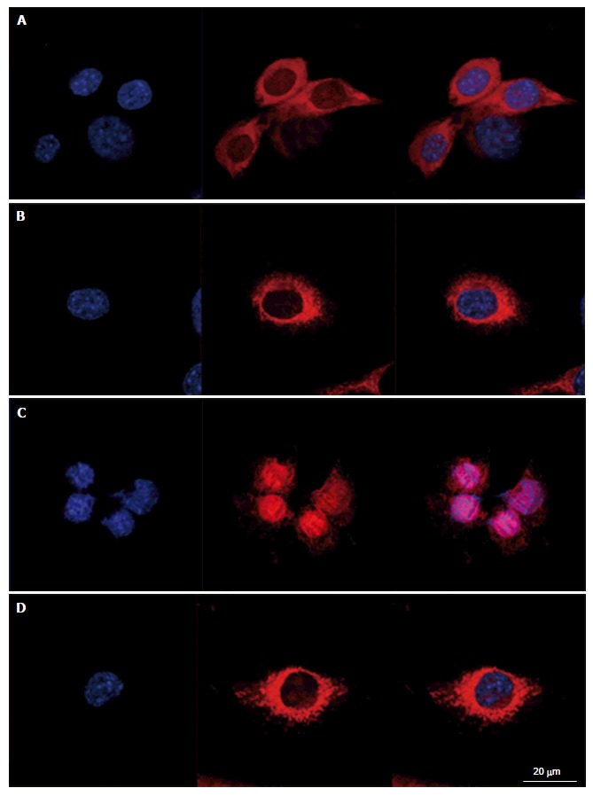
Figure 2.

Prevention of menadione-induced mitochondrial distribution with L-carnitine. C2C12 cells were either untreated or pretreated with 500 μmol/L of L-carnitine and incubated for 24 h with menadione (0 and 9 μmol/L). Nucleus and mitochondria morphology was evaluated after staining with Hoechst 33342 and MitoSoxRed, respectively. From left to right, staining with nuclei, mitochondria and both. Cells mounted in fluorescence medium were observed with a LSM confocal microscope. A: C2C12 untreated with menadione and untreated with L-carnitine; B: C2C12 pretreated with L-carnitine and untreated with menadione; C: C2C12 untreated with L-carnitine and treated with 9 μmol/L of menadione; D: C2C12 pretreated with L-carnitine and treated with 9 μmol/L of menadione.
