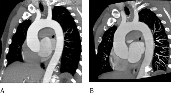
Figure 3.

CT of the thoracic aorta showing moderate dilatation of the ascending aorta. The same patient was studied with the retrospective gated technique with a 320 row-detector, the effective dose was 919 mGy (A). Step and shoot technique used at follow up was able to spare dose (DLP 425 mGy) but misalignment artifacts are evident at the ascending aorta (B).
