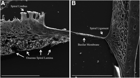
Fig. 2.

Scanning electron microscopy images of decellularized mouse cochlea. The Everhart-Thornley detector (ETD) was used for imaging. In panel a, the medial portions of the cochlea are shown, and the extracellular structures, which include the spiral limbus and osseous spiral lamina are intact; however, no cellular structures are identifiable. Panel b shows the lateral portion of the cochlear duct, in which the basilar membrane and spiral ligament are identified, but the organ of Corti is not present. The extracellular fibers are clearly visible in both panels. Panel a scale bar = 50 μm. Panel b scale bar = 100 μm
