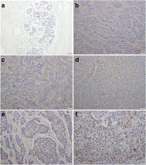
Fig. 1.

Representative breast tissue sections stained with an antibody to EZH2. Representative examples of primary tissue or metastatic tissue cores presenting with five levels of staining for enhancer of zeste homolog 2 (EZH2):a normal breast; b ≤1/100 cells stained (Score 1); c ≤1/10 cells stained (Score 2); d ≤1/3 cells stained (Score 3); e ≤2/3 cells stained (Score 4); and f >2/3 stained (Score 5) (Original magnification, 200× The under bar is 200 μm.)
