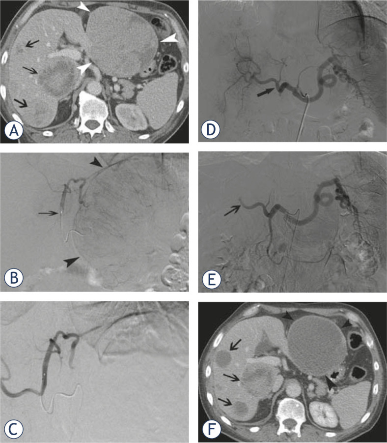
Figure 1.

A 63-year-old male patient presented with a carcinoid of the lung and diffuse bilobar liver involvement. (A) Portal venous phase contrast-enhanced CT-scan confirms diffuse metastatic involvement of both liver lobes (white arrowheads at the level of the largest metastasis in the left liver lobe; black arrows at the level of multiple smaller lesions in the right liver lobe); Selective angiogram of the left hepatic artery (B) before and (C) after chemoembolization with doxorubicin-eluting SAP-microspheres (arrow at the level of the micro-catheter in the left hepatic artery); Selective angiogram of the celiac trunk (D) before and (E) after chemoembolization with doxorubicin-eluting SAP-microspheres (arrow shows stasis of contrast at the level of the right hepatic artery); (F) Portal venous phase contrast-enhanced CT-scan 10 weeks after initial chemoembolization shows marked decrease in volume and enhancement of most of the metastatic lesions in left and right liver lobes.
