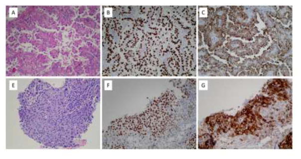
Figure 2.
A. Pulmonary adenocarcinoma, right middle lobe, H&E (200X). B. Pulmonary adenocarcinoma positive for TTF-1 (200X). C. Pulmonary adenocarcinoma positive for Napsin A (200X). Immunohistochemical stains for p63 and CK5/6 are negative (not shown). E. Squamous cell carcinoma, right cardiophrenic angle, H&E (200X). F. Squamous cell carcinoma positive for p63 (200X). G. Squamous cell carcinoma positive for CK5/6 (200X). An immunohistochemical stain for TTF-1 is negative (not shown).

