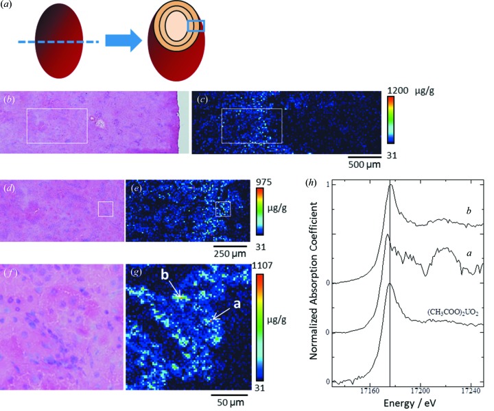
Figure 4.
Uranium distribution in kidney and uranium L III-edge XANES spectra of spots of concentrated uranium in the proximal tubules. The renal section was obtained from an adult rat at the middle phase after uranium exposure at the low dose (day 3 post-injection, 0.5 mg kg−1 body weight). (a) Diagram of the analyzed area of the renal specimen. (b, d, f) Serial-section stained using hematoxylin and eosin. (c) Uranium imaging (150 × 50 steps at 20 µm per step, beam size 1 µm × 1 µm). (e) High-resolution uranium imaging of the boxed area in (b) and (c) (100 × 50 steps at 10 µm per step, beam size 1 µm × 1 µm). (g) High-resolution uranium imaging of the boxed area in (d) and (e) (75 × 75 steps at 2 µm per step, beam size 1 µm × 1 µm). Here, point a indicates the first-highest uranium concentrated spot (1114 µg g−1) and point b indicates the second-highest uranium concentrated spot (948 µg g−1) in the analysed area. The periphery of the renal cortex in all images is shown on the right-hand side. The mean renal uranium concentration was 8.46 µg g−1. (h) Uranium L III-edge XANES spectra of spots of concentrated uranium; graph a is for position a and graph b is for position b in panel (g).

