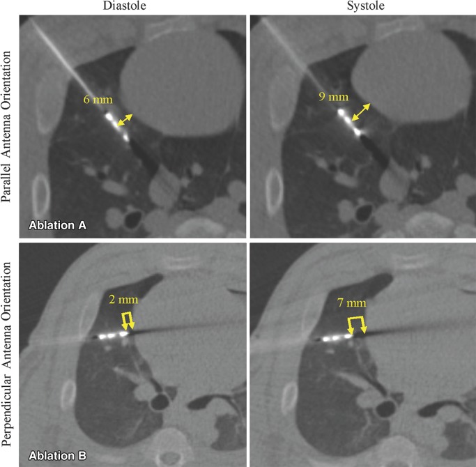
Figure 2:
CT fluoroscopy to record cardiac excursion and to measure antenna distance from heart during diastole. Axial CT fluoroscopic images from ablations performed with, Ablation A, parallel and, Ablation B, perpendicular orientation. Note change in antenna-heart distance from diastole to systole (double arrows). Antenna-heart distance was measured from antenna emission point for parallel ablations and from tip for perpendicular ablations.

