. 2017 Feb 8;58(2):117–126. doi: 10.4111/icu.2017.58.2.117

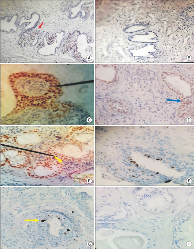
Fig. 2. Immunohistochemical analysis of estrogen receptor (ER) subtypes and Ki-67 expression in prostatic specimens from benign prostatic hyperplasia (BPH) patients (A, C, E, G) and controls (B, D, F, H). (A) Section from BPH specimens shows positive immunostaining (red arrow) for ER beta (ERβ) (H&E, ×40). (B) Representative area from normal prostate shows absence of ERβ immunostaining (H&E, ×40). (C) Intense ERβ positivity was primarily localized to epithelial nuclei (black pointer) in BPH tissues, whereas (D) nuclear staining was sparse (blue arrow) and seen mostly in epithelium of normal prostates (C, D: H&E, ×400). (E) In BPH specimens, immunoreactivity for ER alpha (ERα) was intensely expressed in both epithelial and stromal compartments. Note the close proximity between immunoreactive epithelial and stromal cells (black pointer and yellow arrow) (H&E, ×400). (F) ERα was expressed with much lower intensity in both stromal and epithelial compartments in controls (H&E, ×400). (G) Abundant nuclear staining for Ki-67 (yellow arrow) was found in BPH specimens, whereas (H) a representative area from normal prostate shows absent staining (G, H: H&E, ×400).

Fig. 2