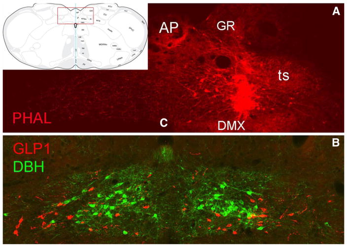
Fig. 1.
PHAL tracer delivery site within the cNST. The inset at upper left shows the approximate rostrocaudal level targeted (~14.2 mm caudal to bregma), and the boxed region corresponds to the photographic images shown in a, b. a A PHAL tracer delivery site (red) centered in the cNST, just dorsal to the DMX. b An adjacent tissue section immunolabeled for GLP1 (red neurons) and DBH (green neurons) to illustrate the locations of intermingling but separate populations of GLP1 and A2 noradrenergic neurons. AP area postrema, c central canal, DBH dopamine beta hydroxylase, DMX dorsal motor nucleus of the vagus, GR gracile nucleus, PHAL phaseolus vulgaris leucoagglutinin, ts tractus solitarius

