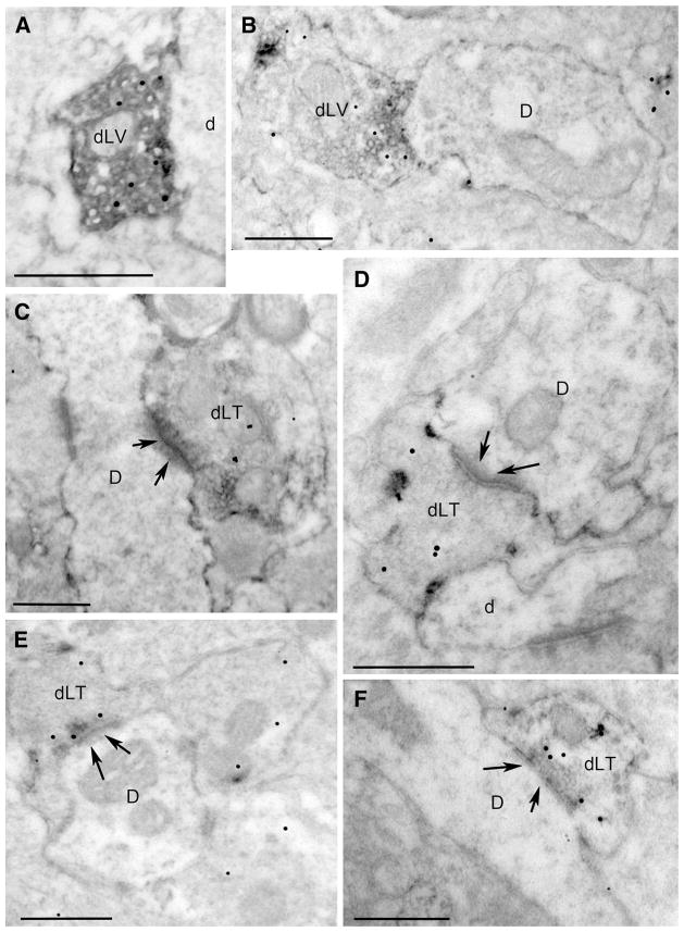
Fig. 5.
Ultrastructural localization of GLP1 immunoperoxidase (floccular electron-dense label) and VGLUT2 immunogold (small particulate label) within the PVN (a–c) and DMH (d–f). D dendrite, dLT double-labeled terminal, dLV double-labeled varicosity. Arrows point out postsynaptic specializations of asymmetric (i.e., excitatory-type) synapses formed by double-labeled axon terminals onto unlabeled dendrites. All scale bars 500 nm

