
Figure 1.
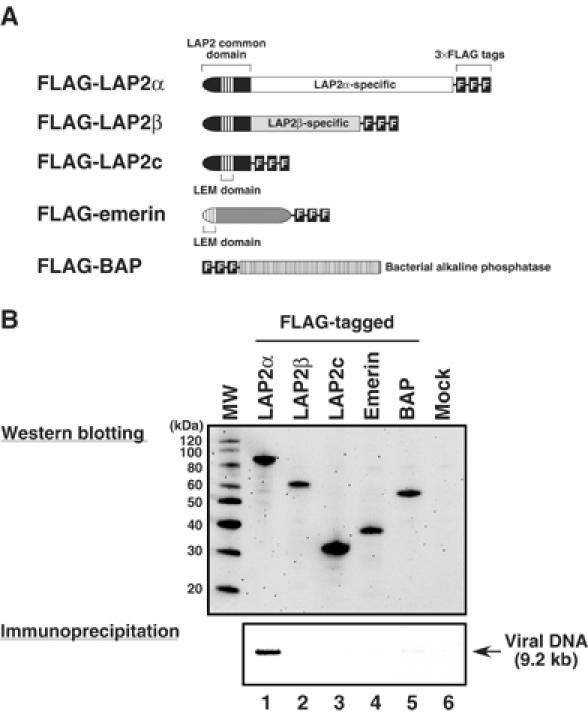
Immunoprecipitation of MoMLV PICs from FLAG-tagged LEM protein-expressing cells. (A) FLAG-tagged LEM protein constructs. (B) Expression of the FLAG-tagged LEM proteins in NIH3T3 cells and immunoprecipitation of PICs from these cells. NIH3T3 cells were transfected with the expression vectors encoding FLAG-tagged proteins and, at 24 h after transfection, the cells were cocultured with MoMLV-producing cells. Cytoplasmic extracts from these cells were immunoprecipitated with an anti-FLAG monoclonal antibody. The weak band in lane 5 of panel B is background that is not reproducibly observed. Viral DNA was extracted from the captured immunocomplex and detected by Southern blotting (lower panel). Protein expression in the cocultured cells was analyzed by western blotting using anti-FLAG monoclonal antibody (upper panel). MW: molecular weight marker. Control experiments demonstrated that the anti-FLAG antibody immunoprecipitates each of the LAP2 isoforms with similar efficiency (Supplementary Figure S1).