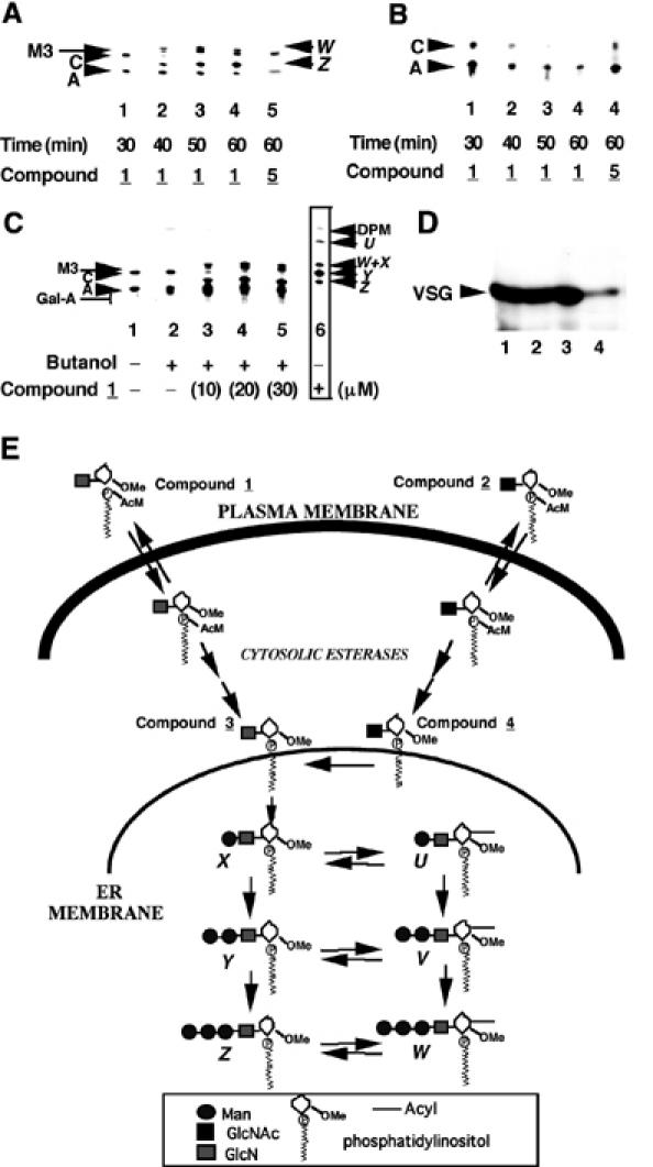
Figure 2.

Compound 1 is metabolized by the trypanosome GPI pathway in vivo and inhibits the formation of glycolipid C. (A) Living trypanosomes were pre-incubated with 50 μM compound 1 or compound 5 for 30–60 min, as indicated, and then labelled for 60 min with [3H]Man. Extracted glycolipids were analysed by HPTLC and fluorography. The products, which were characterized by enzymatic and chemical digestions (Table I), are as described in the legend to Figure 1. (B) As for (A), but labelling with [3H]myristate. (C) As for (A), but with a fixed pre-incubation time of 60 min and varying concentrations of compound 1. Compound 1 was added as a butanol solution and a control with butanol alone is included (lane 2). The boxed data (lane 6) are from a cell-free system labelling experiment to provide markers for glycolipids U–Z. (D) Trypanosomes were pre-incubated without any additive (lane 1), or with 30 μM compound 1 (lane 2), 30 μM compound 2 (lane 3) or 60 μg/ml cycloheximide (lane 4) for 60 min, labelled with [3H]myristate for 60 min, lysed in boiling sample buffer and analysed by SDS–PAGE and fluorography. The radiolabel incorporated in the presence of the protein synthesis inhibitor cycloheximide represents that due to myristate exchange on pre-existing VSG rather than de novo synthesis of VSG. (E) Summary of the fates of compounds 1 and 2, as indicated by the in vivo labelling data (see text).
