
Figure 6.
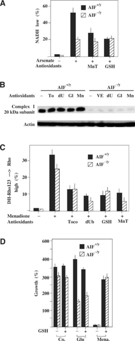
Modulation of the redox consequences of the AIF defect. (A) Enhanced NAD(P)H depletion in AIF+/y as compared to AIF−/y cells in response to arsenate. Cells were treated with 1 mM arsenate for 6 h, in the presence or absence of GSH ester (5 mM) or MnTBAP (50 μM), followed by cytofluorometric determination of cellular NAD(P)H levels. (B, C) Failure of antioxidants to cause re-expression of the 20 kDa complex I subunit. AIF−/y and AIF+/y cells were cultured for 72 h in the presence of tocopherol (200 μM), decylubiquinone (50 μM), GSH ester (10 mM), or MnTBAP (50 μM), all re-added every 12 h, followed by immunoblot (B). Alternatively, cells were treated for 3 h with menadione (100 μM) and the production of ROS was measured by assessing the oxidation of dihydrorhodamine 123 in the cytofluorometer (C). (D) Failure of the antioxidant GSH to rescue AIF−/y cells from glucose withdrawal-induced cell death. AIF−/y and AIF+/y cells were cultured for 48 h in medium without glucose and/or 10 mM GSH ethyl ester, followed by determination of viability as in Figure 5C. As an internal control of the GSH effect, AIF+/y cells were also cultured in the presence of menadione (100 μM). The values are means±s.e.m. of three independent determinations.