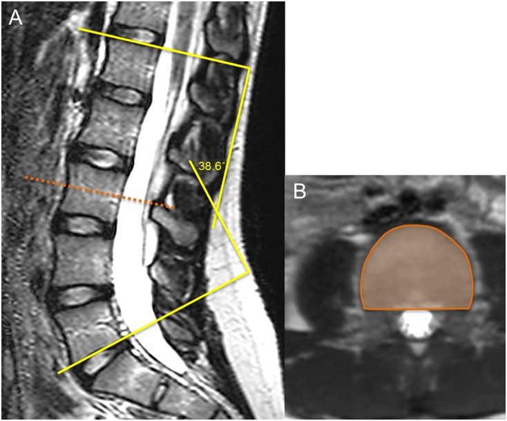
Fig 1. MRI images of the lumbar spine in a 13-year-old girl.
(A) Coned-down sagittal image of the lumbar spine showing the degree of lumbar lordosis measured as the angle between the superior endplate of L1 and the inferior endplate of L5. (B) Axial image outlining the measurement of vertebral CSA at the third lumbar vertebra.

