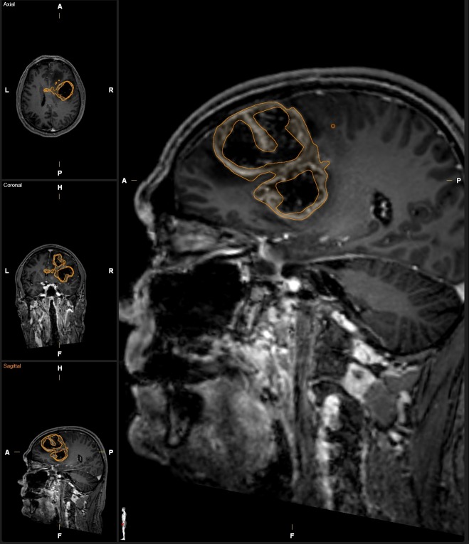
Fig 1. Example segmentation of a complex GB.
Contrast enhancing tumor volume was delineated on a contrast enhanced MPRage image set rendering a complex tumor shape. Necrotic areas were avoided during segmentation as it was feasible. The small spot located posterior to the segmentation on the magnified sagittal image represents the “brush” of the semi-automated region growing tool.

