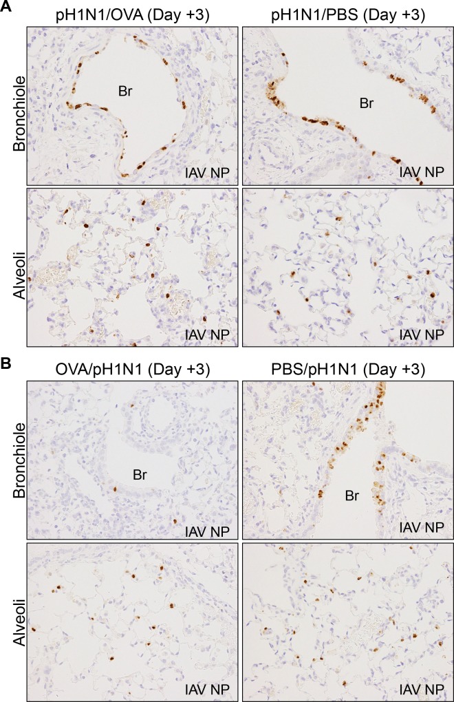
Fig 4. Immunohistochemical analysis of influenza A virus NP antigen (IAV NP) expression in the bronchiolar and alveoli epithelium of pH1N1-infected mice at Day 3 post-infection.
(A) Representative sections of lung tissue from mice infected with influenza virus followed by induction of allergic airway responses using OVA (pH1N1/OVA) or from mice infected with influenza and subsequently challenged with PBS (pH1N1/PBS). (B) Representative sections of lung tissue from the mice infected with influenza virus and pre-existing OVA-induced s inflammation (OVA/pH1N1) and from mice infected with influenza virus in the absence of allergic airway inflammation (PBS/pH1N1) The data are representative of 3 mice/group from two independent experiments. Original magnification: ×40. Br, Bronchiole.

