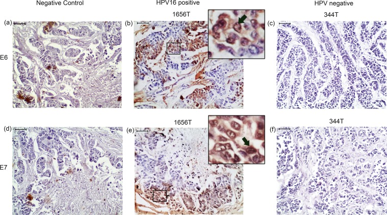
Fig 6. Immunohistochemical detection of E6 and E7 protein of HPV16 in pre-therapeutic breast tumor tissues.
(b) & (e) Representative immunohistochemical staining of E6 and E7 in HPV16 positive samples. (c) & (f) Representative immunohistochemical staining in HPV negative sample (a) & (d) Immunohistochemical staining with out primary antibody represented Negative control (NC). [Magnification of tissue samples is 20X and for inset, magnification is 40X, Scale bars = 50 μm].

