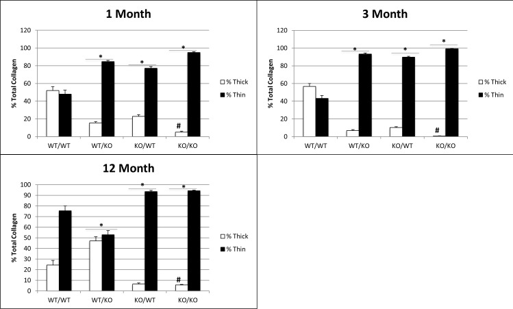
Fig 2. A consistent reduction in collagen I fiber size observed in double-null PDL from 1-month to 12-months of age.
Collagen fiber morphology represented by thin vs. thick fibers quantified from PSR histology for three age points. n = 5 mice/genotype, 5 sections/mouse. *p<0.05 compared to wt/wt counterparts, #p<0.05 compared to single transgenics.

