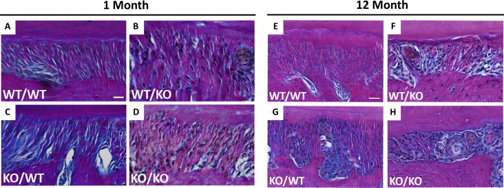
Fig 3. Collagen fiber morphology altered in double-null PDL at 1- and 12-month age points.
Representative images of Herovici stained PDL from wt/wt (A&E), wt/ko (B&F), ko/wt (C&G), and ko/ko (D&H) shown at 1- and 12-months of age respectively. Red/pink stain indicates thicker collagen fibers; blue stain indicates thinner collagen fibers, nuclei stain black. Images captured at 40X and oriented with alveolar bone on bottom, PDL center, and tooth cementum on top. Bar in A&E = 25 μm and applies to all panels. n = 3 mice/genotype, 5 section/mouse.

