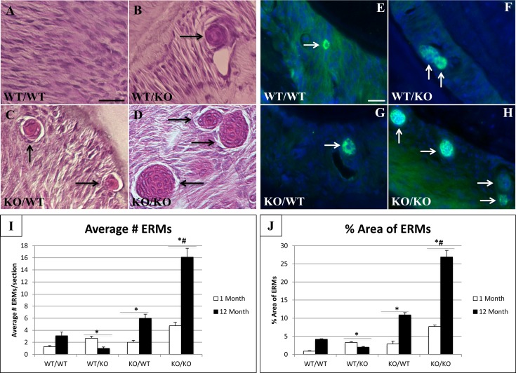
Fig 6. Number and size of epithelial rests of Malassez (ERMs) varies in murine periodontal ligament (PDL).
Representative sections from 1 month wt/wt (A), wt/ko (B), ko/wt (C), and ko/ko (D) PDL stained with H&E. Black arrows indicate multi-cellular ERMs. Bar in A = 25 μm and applies to all panels. Representative images from 1 month PDL stained for cytokeratin immunoreactivity (green); wt/wt (E), wt/ko (F), ko/wt (G), ko/ko (H). Nuclei stained blue (DAPI). White arrows indicate multi-cellular rests of Malassez expressing cytokeratin. Images are representative of n = 3 mice/genotype. I. Quantification of average number of rests present in sampled PDL. J. Percent of PDL area occupied by rests. n = 5. *p<0.05 compared to wt/wt, #p<0.05 compared to single transgenics.

