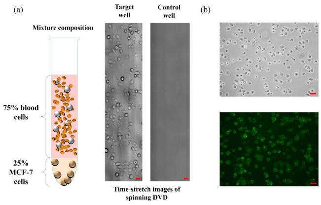
Fig. 5.
(a) (Left) Specimen of MCF-7 (25%) mixed with human buffy coat (75%) used for antibody-captured, and thus enrichment of, MCF-7. (Middle) Time-stretch image of the enriched MCF-7 (with anti-EpCAM antibody) in the target well. (Right) Time-stretch image of the control well in which only streptavidin is coated. Both the images in (a) were taken at a spinning speed of 900 rpm (linear speed of 4 m/s). Same substrate design as shown in Fig. 4. was adopted in this experiment. (b) Static images (Top: phase contrast; Bottom: fluorescence) of the enriched MCF-7 further stained with green fluorescent dye (Alexa Fluor-488 anti-EpCAM (RnD FAB9601G, 10 µg/mL)). This additional staining step was performed to further confirm the MCF-7 enriched on the DVD. All scale bars (in red) represent 50 µm.

