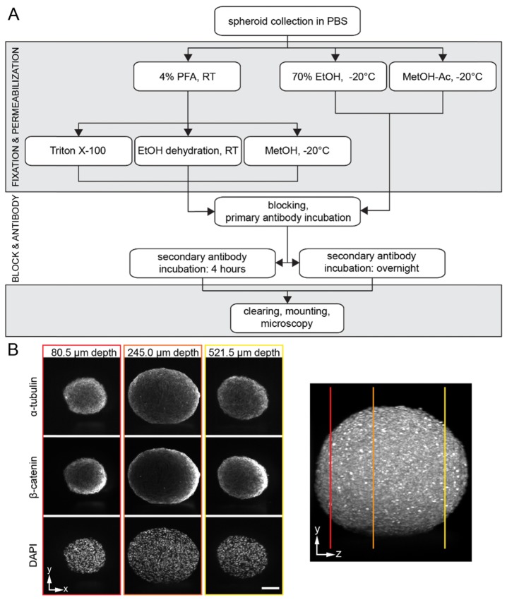
Fig. 1.
(A) Schematic illustration of the immunostaining protocols. The workflow shows the different steps of the immunofluorescence staining starting with the collection of the spheroids after six days of formation. The main steps are fixation, permeabilization, blocking, antibody incubation and optical clearing. (B) Single planes at different depths in a typical spheroid data set are shown. The right image shows a 90° rotation around the y-axis of the data set that depicts the location of the single planes. Microscope: mDSLM; illumination objective: Epiplan-Neofluar 2.5x/NA 0.06; detection objective: N-Achroplan 10x/NA 0.3; α-tubulin: 561 nm, 0.09 mW, bandpass filter 607/70; β-catenin: 488 nm, 0.134 mW, bandpass filter 525/50; DAPI: 405 nm, 0.01 mW, bandpass filter 447/55; scale bar: 100 µm. h: hours, min: minutes, RT: room temperature.

