Fig. 2.

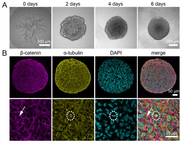
Immunostainings against α-tubulin and β-catenin in histological sections of U343 spheroids show a homogeneous signal throughout the whole spheroid. (A) U343 spheroids were formed for six days from 10,000 seeded cells. (B) Paraffin-embedded spheroids were cut into 4 µm thick sections and stained against α-tubulin and β-catenin. A section from the middle part of a spheroid shows a homogeneous distribution of the signal for α-tubulin as well as β-catenin. Cell nuclei were counterstained with DAPI. β-catenin is located at the plasma membranes of cells (arrow). The α-tubulin antibody specifically labels the microtubules in U343 spheroids, which is best seen in the mitotic spindle during cell division (dashed circle). Microscope: Zeiss LSM780; objective lens (upper panel): Plan-Apochromat 20x/numerical aperture (NA) 0.8; objective lens (lower panel): Plan-Neofluar 40x/NA 1.3.
