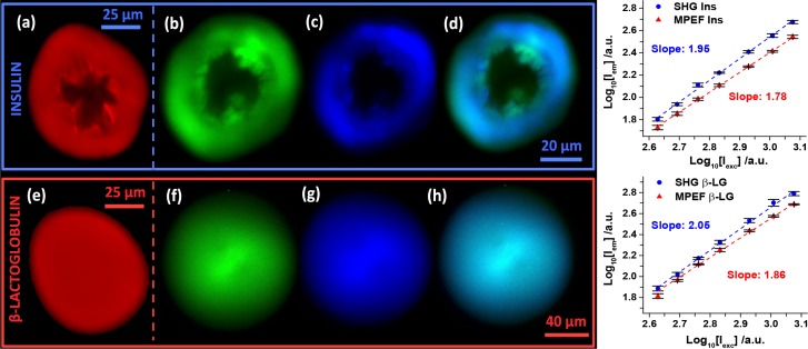
Fig. 3.
Label-free imaging of amyloid spherulites. Spherulites from bovine insulin (a–d) and β-LG from bovine milk (e–h) imaged with confocal fluorescence (a,e - red), MPEF (b,f - green), and SHG (c,g - blue). MPEF and SHG were imaged simultaneously in different channels (420–460 nm and 495–540 nm, respectively) and overlays of the two are shown (d,h). Scale bars: 25 μm (a,e), 20 μm (b–d), and 40 μm (f–h). The λexc were 405 nm (a,e) and 910 nm (b–d,f–h). The power dependences for SHG and MPEF were measured for four replicates of both structures with powers ranging from 425–1190 mW. The error bars represent the St. Dev. and the slopes were obtained with least square fits.

