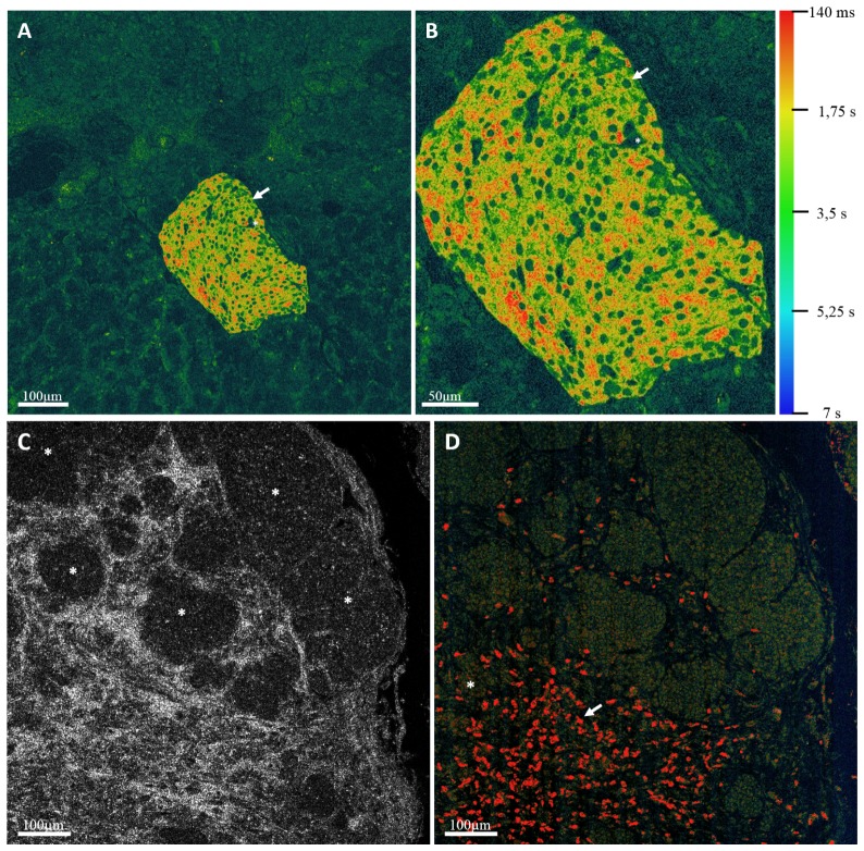
Fig. 9.
(A) shows a Dynamic FF-OCM image of a Langerhans islet inside a fresh ex-vivo rat pancreas. One can notice the nuclei (arrow) with a circular shape and capillaries (*) irrigating the islet. (B) is a detailed view of the islet. (C) Shows the FF-OCM image a mouse intestinal tumor where the collagen matrix is highly visible compared to the cells inside the nests (*). (D) is a Dynamic FF-OCM image of the same field as (C), the collagen fibers are no longer visible because they are stationary, whereas the cancerous cells appear inside the nests and immune cells are revealed inside the collagen matrix (arrow). We also remove the ambiguity on zones like (*) and below where it was not clear if cancerous cells were present. The color bar is the same for the 3 images.

