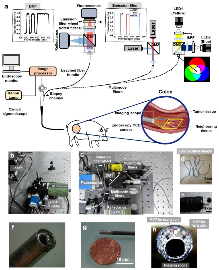
Fig. 1.
Configuration of the clinically-compatible flexible wide-field multi-color fluorescence endoscope system. (a) Schematic of multi-color fluorescence endoscope designed to be inserted through the biopsy channel (diameter: 3.2 mm) of a clinical sigmoidoscope. The system is comprised of two detection channels: reflectance and fluorescence. Multiple-channel fluorescence imaging is obtained by the rotating filter wheel in front of the fluorescence camera detector. (b–c) Photographs of the imaging device and system. (c) An enlarged view of the detection part in the endoscopic system. (d) Photograph of highly flexible leached fiber bundle used for home-made probes. Note that the leached fiber bundle in (a) is not visible in (c). (e) Photograph of combined imaging scope and sigmoidoscope. (f–g) Close-up photograph of the distal end of the imaging scope. The scope was enclosed and sealed within an aluminum sheath. The magnified imaging scope shows a leached fiber bundle and multimode fibers for delivering excitation light and white light (from LEDs). (h) Frontal view of the distal end of the imaging scope. Acronyms are defined as follows: (LED: Light-emitting diode, DM: Dichroic mirror, MMF: Multi-mode fiber, BPF: Band-pass filter, RGB: Red green blue).

