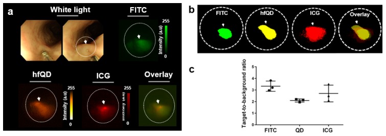
Fig. 6.
In vivo multi-color fluorescence endoscopic imaging in the porcine colon. (a) (upper) Generation of a surrogate tumor with mixtures of multiple fluorophores. Fluorescence images of each dye were obtained with corresponding emission filters. (b) Threshold images of multiple fluorophores with the endoscope showing the distributions of each fluorophore and a merged image. (c) TBR of fluorophores in the surrogate tumor at the same concentration of 1 mg/ml.

