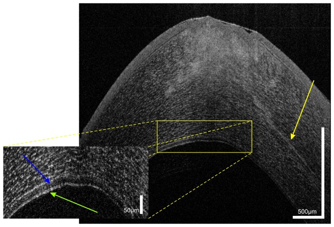
Fig. 8.
UHR-OCT of Acanthamoeba keratitis in the same patient as shown in Fig. 7. The cross-sectional image reveals radial keratoneuritis with a thickened corneal nerve and a hyporeflective space (blue arrow) above the double banded structure of the epithelium.

