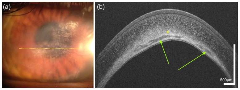
Fig. 9.
Sequelae after herpetic keratitis in a 34 year old male who was first diagnosed 3 years ago. The slit lamp photography (a) as well as the OCT image (b) reveal neovascularization. Yellow dotted line in (a) indicates the location of the UHR-OCT scan; Green arrows, corneal neovascularization; yellow asterisk, calcifications/lipid.

