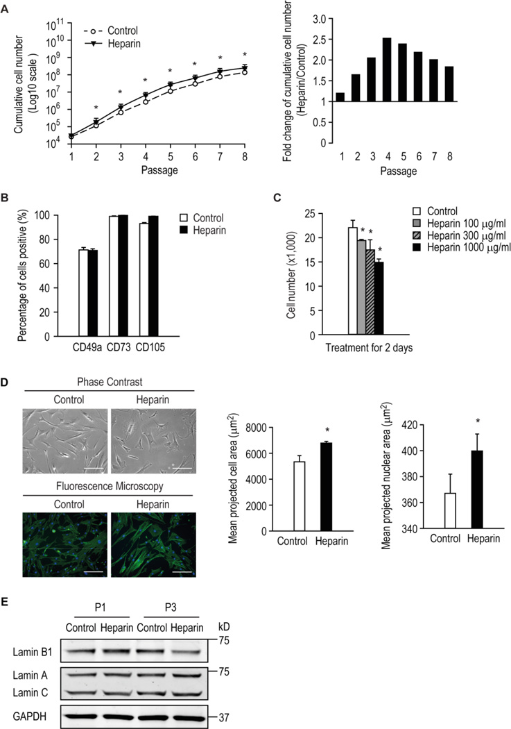
Figure 1. The response of hMSCs to various doses of heparin.
Human MSCs were serially passaged in the presence or absence of 160 ng/ml heparin and cumulative growth assessed (A) and the expression of surface antigens common to hMSCs by flow cytometry at late passage (B). * p < 0.05 versus control. (C) Number of hMSCs cultured with or without heparin at the indicated doses for 2 days. (D) left panel, hMSCs were treated for 3 days with or without 500 µg/ml heparin and imaged using phase contrast or fluorescence microscopy after cells were stained with DAPI and phalloidin to visualize nuclei in blue and actin cytoskeleton in green, respectively; right panel, image cytometric quantification of cell and nuclear projected area. * p < 0.05 versus control. (E) Human MSCs were grown in the presence or absence of 500 µg/ml heparin for one (P1) or three (P3) passages. The levels of target proteins were detected by Western blot analysis.

