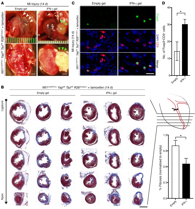
Figure 7. Hydrogel treatment with IFN-γ after MI rescues the mutant phenotype.
(A) Whole-mount bright-field images of representative Wt1CreERT2/+ Yapfl/fl Tazfl/fl R26Tomato/+ (mutant) hearts following MI injury with empty or IFN-γ gel treatment. White arrowheads indicate adhesion of the anterior heart to the chest wall; green arrowheads denote hydrogel on the heart surface. (B) Masson’s trichrome staining of serial cross sections from the coronary ligature to the apex of the heart 14 days after MI in empty gel– and IFN-γ gel–treated animals (n = 4 for each cohort). Fibrotic tissue is stained blue. Quantitation of the proportion of fibrosis indexed to the total myocardial CSA in the LV free wall revealed a 34.4% relative reduction of fibrosis in IFN-γ–treated hearts compared with empty gel–treated hearts (P < 0.05). (C) Cross sections were immunostained as indicated. (D) Mean number of CD3+Foxp3+ cells per heart section in 4 mice per group. Scale bars: 1 mm (A), 2 mm (B), and 20 μm (C). Data represent the mean ± SEM. *P < 0.05, by 2-tailed Student’s t test.

