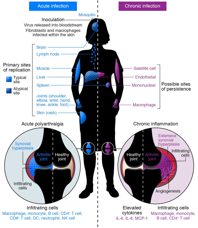
Figure 3. Model of acute and chronic CHIKV pathogenesis.
Acute CHIKV infection begins with transmission of the virus via a bite of an infected mosquito to the skin, where it replicates in susceptible cells, including fibroblasts and macrophages. The virus disseminates through lymphatics and bloodstream to typical (solid) and atypical (hatched) sites of primary replication (indicated in blue). Acute infection elicits an inflammatory response in infected tissues, characterized by an extensive infiltration of mainly macrophages and monocytes, but also neutrophils, NK cells, and lymphocytes in target tissues (indicated in blue), and by elaboration of a number of proinflammatory chemokines and cytokines. Within arthroskeletal tissues, synovial hyperplasia begins. Viral replication and host immune responses cause myalgia and polyarthralgia in distal joints. Chronic CHIKV disease can persist for months or years after acute infection but is often limited to more distal joints. Chronic disease is likely mediated by persistent virus and inflammation. Possible sites of CHIKV persistence include endothelial cells in the liver and other organs, mononuclear cells in the spleen, macrophages within the synovial fluid and surrounding tissues, and satellite cells within the muscle (indicated in purple). Within the chronically infected joint, the continued presence of a subset of infiltrating cells (mainly macrophages, monocytes, and lymphocytes) and specific proinflammatory mediators (IL-6, IL-8, and MCP-1) within the synovial fluid likely contribute to prolonged inflammatory disease. Chronic joint pathology resembles that in rheumatoid arthritis, with significant hyperplasia and angiogenesis. This model is based on human and animal studies.

