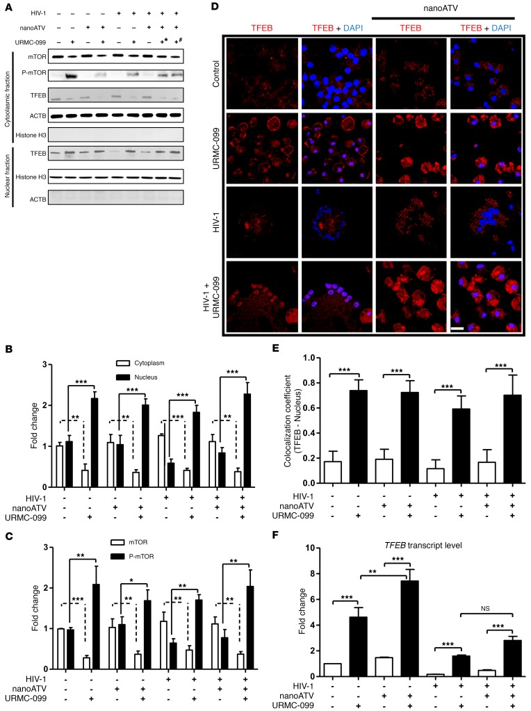
Figure 2. URMC-099 regulates TFEB nuclear localization.
Human MDMs were treated for 14 days with 100 (+*) or 400 ng/ml (+#) URMC-099 in the presence or absence of 100 μM nanoATV, with or without HIV-1ADA infection. (A) MDMs were fractionated and analyzed by Western blotting. (B) TFEB and (C) mTOR quantification of Western blots. Protein bands were quantified and normalized to actin β (ACTB) or histone H3 using ImageJ2 software. (D) Human MDMs were stained for TFEB (red) and DAPI (blue) and analyzed using a confocal microscope to visualize nuclear localization of TFEB. Scale bar: 20 μm. (E) Quantification of colocalization coefficient for TFEB and DAPI. (F) Total RNA was isolated on day 14, and real-time qPCR was performed to determine TFEB expression. Values represent the mean ± SD (n = 3). *P ≤ 0.05, **P ≤ 0.01, and ***P ≤ 0.001, by Student’s t test. (B, C, E, and F) Data were corrected for multiple comparisons using the Benjamini-Hochberg method. Data are representative of 3 independent experiments. All URMC-099 treatments not specified used 400 ng/ml.

