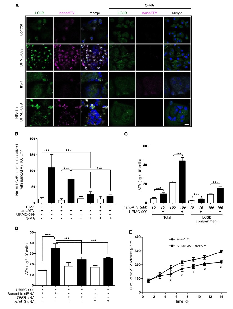
Figure 4. URMC-099 increases nanoATV retention in macrophage autophagosomes.
(A) Human MDMs were treated for 14 days with 400 ng/ml URMC-099 in the presence of fluorescently tagged nanoATV (10 μM) (purple), with or without HIV-1ADA infection. MDMs were transfected with an LC3B-tagged GFP construct (green), stained with DAPI (blue), and analyzed by confocal microscopy. Scale bar: 20 μm. (B) Quantification of LC3B puncta that colocalized with nanoATV. (C) Human MDMs were treated with nanoATV (10 μM or 100 μM) in the presence or absence of 400 ng/ml URMC-099. After 14 days, the ATV concentration was determined in total cells, and LC3B autophagosomal compartments were isolated using magnetic bead separation (n = 3). (D) Human MDMs were treated with 100 μM nanoART and 400 ng/ml URMC-099. Cells were transfected with siRNA for TFEB or ATG13, and on day 14, the ATV concentration was determined in total cells (n = 3). (E) URMC-099 regulated the release of nanoATV. Human MDMs were treated with 100 μM nanoATV for 16 hours, washed with PBS, and incubated with or without 400 ng/ml URMC-099. On different days, supernatant was collected, ATV concentration was quantified using HPLC, and the cumulative ATV release was plotted according to treatment duration (n = 3). Comparison of the means by 2-way factorial ANOVA showed a time-dependent treatment effect (P = 0.0002), with pairwise comparisons made using Bonferroni’s post-hoc test (#P < 0.05). Data are representative of 3 independent experiments. (B–D) Values represent the mean ± SD. *P ≤ 0.05 and ***P ≤ 0.001, by Student’s t test. Multiple comparisons were corrected for the FDR using the Benjamini-Hochberg method.

