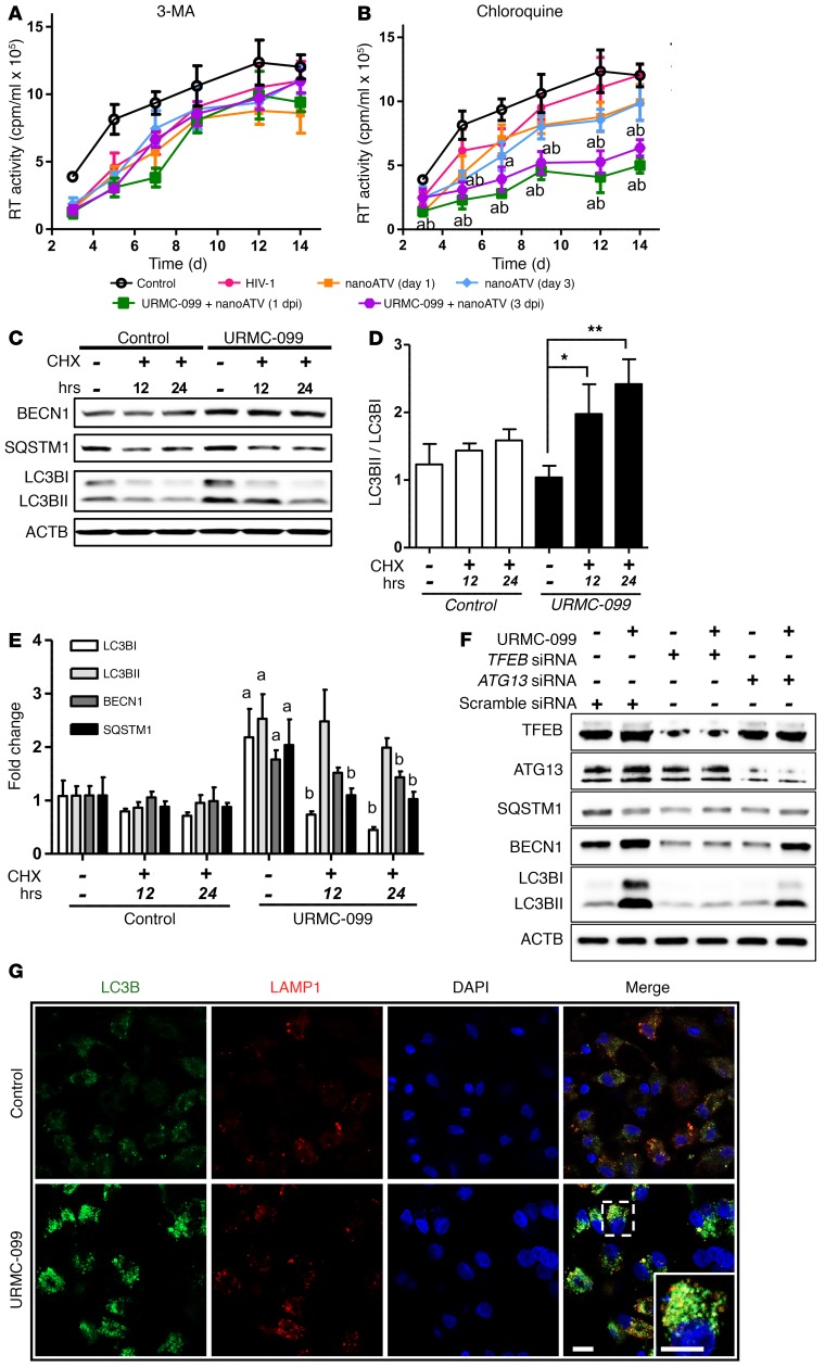
Figure 5. URMC-099–induced autophagy affects HIV-1 clearance.
HIV-1ADA–infected human MDMs were treated with 1 μM nanoATV on day 1 or day 3 after infection and incubated with or without 400 ng/ml URMC-099 and in the presence of the autophagy inhibitors (A) 3-MA (100 μM) or (B) chloroquine (10 μM). Supernatants were collected on different days after infection and analyzed for HIV-1 RT activity (n = 5). The same HIV-1 infection control plot is presented in A and B. (A and B) The mean values of RT activity were assessed by 2-way factorial ANOVA, which showed a significant time-dependent treatment effect (P < 0.02). Pairwise comparisons using Bonferroni’s post-hoc test were assessed for URMC-099–treated cultures, with P < 0.05 compared with HIV-infected controls in the absence (acontrol) or presence (bHIV) of an autophagy inhibitor. (C–E) MDMs were treated in the presence or absence of 400 ng/ml URMC-099 for 14 days. Twenty-four hours or twelve hours before harvesting, cells were treated with 10 μM cycloheximide (CHX) to inhibit translation. Total cell lysates were analyzed by Western blotting. (D) Values represent the mean ± SEM of LC3BII/LC3BI ratios and were compared by Student’s t test and adjusted for multiple comparisons using the Benjamini-Hochberg method. *P ≤ 0.05 and **P ≤ 0.01 (n = 3). (E) Differences in mean fold changes were assessed by 2-way ANOVA and pairwise comparison with the respective proteins was done using Bonferroni’s post-hoc test. P ≤ 0.05 for ano URMC-099/no CHX control and bURMC-099/no CHX control (n = 3). (F) Human MDMs treated with 400 ng/ml URMC-099 were transfected with either TFEB siRNA or ATG13 siRNA on days 3 and 7. On day 14, cell lysates were analyzed for by Western blotting. (G) URMC-099–treated (400 ng/ml) and untreated (control) MDMs were transfected with LC3B-GFP on day 12, and 48 hours later were stained and imaged with a confocal microscope. Scale bars: 20 μm. Data are representative of 3 independent experiments.

