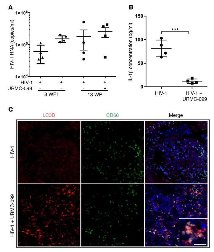
Figure 7. URMC-099 treatment in humanized mice.
(A and B) Humanized NSG mice were infected with HIV-1ADA and treated with URMC-099 (n = 4 per group). Ten weeks after infection, (A) plasma viral load and (B) plasma IL-1β concentration were determined (n = 4 mice per group). Values represent the mean ± SEM. ***P ≤ 0.001, by Mann–Whitney U test. (C) Paraffin-embedded spleen tissue sections from URMC-099–treated and control HIV-1–infected humanized mice were stained for LC3B (red) in autophagosomes and CD68 (green) in macrophages. DAPI was used to stain nuclei. The merged panel shows colocalization of LC3B and CD68 in spleens from URMC-099–treated mice. Scale bars: 20 μm.

