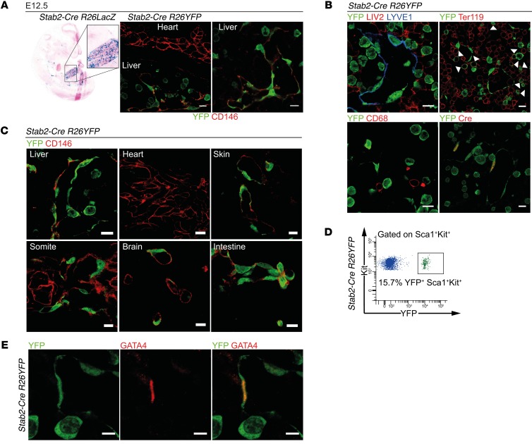
Figure 2. Characterization of endothelial subtype–specific Stab2-Cre driver mice.
(A) Analysis of Stab2-Cre R26LacZ (β-gal assay) (n = 3) and Stab2-Cre R26YFP (co-IF of YFP and CD146) (n = 3) shows reporter activity in the endothelium of the fetal liver. Scale bars: 10 μm. (B–E) Analysis of the Stab2-Cre R26YFP reporter in the embryo. (B) Co-IF of YFP and LYVE1, Cre, LIV2, Ter119, and CD68 in the fetal liver at E12.5. Arrowheads indicate YFP+Ter119+ cells (n = 3). Scale bars: 10 μm. (C) Co-IF of YFP and CD146 at E12.5 (n = 3). Scale bars: 10 μm. (D) FACS analysis of the fetal liver of Stab2-Cre R26YFP reporter mice at E13.25. Representative FACS blot showing YFP reporter activity in Sca1+Kit+ cells (n = 5). (E) Co-IF of YFP and GATA4 in the fetal liver at E12.5 (n = 3). Scale bars: 5 μm.

