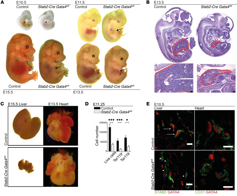
Figure 3. Deletion of GATA4 in LSECs in Stab2-Cre Gata4fl/fl mice impairs liver development and causes embryonic lethality.
(A) Photomicrographs of Stab2-Cre Gata4fl/fl embryos at different ages. Arrows indicate hypoplastic livers in the mutant embryos from E11.5 to E15.5 (n > 3). (B) H&E staining of Stab2-Cre Gata4fl/fl embryos at E13.5. Red dotted lines indicate the fetal liver (n = 5). Scale bars: 100 μm. (C) Photomicrographs of the fetal liver (E15.5) and fetal heart (E13.5) of Stab2-Cre Gata4fl/fl embryos. (D) FACS analysis of live cells, Ter119+ cells, and Ter119– cells in the liver of Stab2-Cre Gata4fl/fl embryos at E11.25 (n = 14 mutants and 10 controls). Student’s t test; *P < 0.05, ***P < 0.001. (E) Co-IF of GATA4 and STAB2 or CD31 of fetal livers of Stab2-Cre Gata4fl/fl embryos at E10.5. IF shows absence of GATA4 in the mutant liver (arrowheads), but not in controls (arrows) (n = 3). Scale bars: 10 μm.

