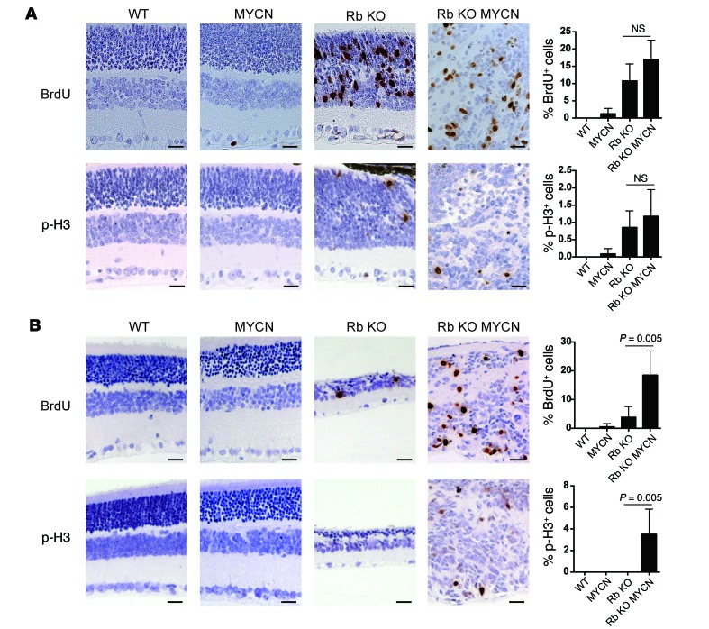
Figure 2. MYCN cooperates with Rb inactivation to increase proliferation in the retina.
(A) Brdu and p-H3 analysis of retinae at P12 showing increased proliferation in Rb-null (Rb KO) and Rb/TET-MYCN (Rb KO MYCN) retinae relative to control or MYCN-only retinae. N = 4–7 independent retinae, with 1 section examined per retina. (B) BRDU and p-H3 analysis at P22 shows increased proliferation in Rb/TET-MYCN retinae relative to Rb-deficient retinae that had largely exited the cell cycle. N = 4–6 independent retinae, with 1 section per retina. Scale bar: 25 μm. Error bars represent the SD. P values shown in B were determined by 2-tailed Student’s t test.

