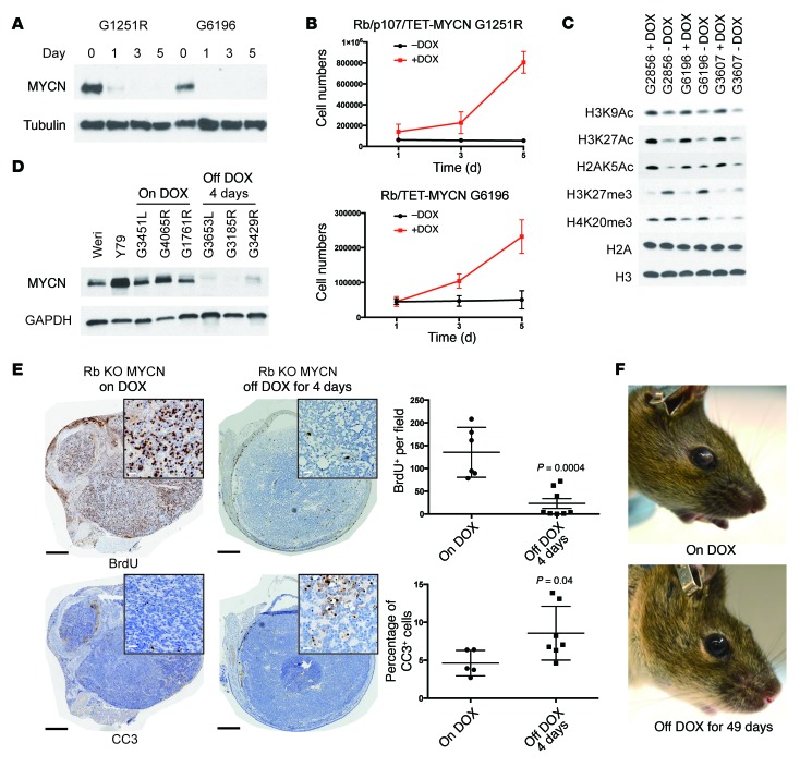
Figure 4. Sustained MYCN expression is initially required for retinoblastoma cell proliferation.
DOX removal in cell lines derived from Rb/TET-MYCN tumors led to (A) reduced MYCN protein expression on Western blotting and (B) proliferation impairment, as assessed by counting cells 1, 3, and 5 days after plating. Data were pooled from 3 independent experiments. (C) Western blot of histone extracts from 3 Rb/TET-MYCN cell lines showing reduced histone H3K9, H3K27, and H2A K5 acetylation (Ac) and increased H3K27 and H4K20 trimethylation (me3) upon removal of DOX from the media. Individual blots indicating equal loading are shown Supplemental Figure 4D. (D) DOX removal in vivo initially resulted in suppression of MYCN protein expression by day 4. (E) Immunohistochemical analysis shows decreased BrdU-positive cells and increased cleaved caspase 3 (CC3) levels 4 days after DOX removal. Black scale bars: 500 μm; white scale bars (insets): 25 μm. Plot shows quantification from 5 to 8 independent retinae per genotype. The P values were determined by Student's t test. (F) Photographs showing a mouse with retinoblastoma in the anterior chamber, compared with 49 days later when the tumor had regressed.

